Areas of The Brain: Definition, Function, & Development
Discover and dive into the different areas of the brain.
*This page may include affiliate links; that means we earn from qualifying purchases of products. |
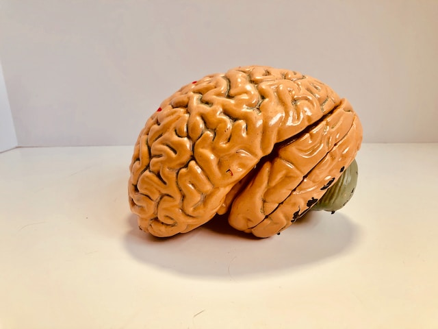
Brains are extraordinarily complicated organs. Even though the average adult human brain weighs about 2.7 lbs and is only about the size of two clenched fists, it contains about 171 billion cells. All of these cells are arranged in a complex topography of hills, valleys, and distinct layers, all contributing to the many remarkable brain functions in their own way. |
Before reading on, if you’re a therapist, coach, or wellness entrepreneur, be sure to grab our free
Wellness Business Growth eBook to get expert tips and free resources that will help you grow your business exponentially.Are You a Therapist, Coach, or Wellness Entrepreneur?
Grab Our Free eBook to Learn How to
Grow Your Wellness Business Exponentially!
✓ Save hundreds of hours of time ✓ Earn more $ faster
✓ Boost your credibility ✓ Deliver high-impact content
What Are Areas of The Brain? (A Definition)
There are many different levels of organization in the brain. For example, the brain can be first divided into three major parts: the forebrain, the midbrain, and the hindbrain. Each of these three parts can be further divided into several smaller regions, each with its own specific function.
Anatomical regions of the brain are defined based on their location and structure. The boundaries between different regions are typically defined by physical landmarks, such as fissures, sulci, and gyri (the hills and valleys you see when you look at a typical human brain), as well as by the location of specific nuclei and fiber tracts.
One of the most common methods of defining the different regions of the brain is through the use of neuroimaging techniques, such as magnetic resonance imaging (MRI) and computed tomography (CT) scans. These techniques allow researchers to non-invasively visualize the structure and function of the human brain.
Another way to define brain regions is through post-mortem dissection and histological analysis. In this approach, the brain is examined under a microscope, and regions are identified based on their cellular and anatomical characteristics.
In recent years, advances in techniques such as diffusion tensor imaging (DTI) and functional magnetic resonance imaging (fMRI) have allowed researchers to define brain regions based on their connectivity and functional activity. By tracing the connections between different brain regions, scientists can create maps of functional networks in the brain, providing insights into how different areas of the brain interact to support various cognitive and behavioral functions.
Why Are Areas of The Brain Important?

Areas of the Brain and Function
The brain can be divided into regions based on both anatomical landmarks, like specific folds and grooves, as well as based on their different functions. Different regions of the brain have evolved to specialize in different cognitive, motor, and sensory functions, which allows us to describe these regions based on what they do. For example, because vision is processed in a region at the back of your brain, we call this area the visual cortex. We can also refer to other functionally defined regions such as the motor cortex, sensory cortex, and associative cortex.
Areas of the Cerebral Cortex
Frontal lobe
The frontal lobe is located at the front of the brain and is involved in higher cognitive functions, such as reasoning, planning, decision-making, and voluntary movement. It includes the prefrontal cortex, which is involved in executive functions, and the motor cortex, which controls voluntary movement.
Parietal lobe
The parietal lobe is located at the top and back of the brain and is involved in processing sensory information, such as touch, temperature, and pain. It includes the somatosensory cortex, which processes tactile and proprioceptive information, and the association cortex, which integrates information from multiple sensory modalities.
Temporal lobe
The temporal lobe is located on the sides of the brain and is involved in processing auditory information, as well as some aspects of visual perception and memory. It includes the primary auditory cortex, which processes sound, and the hippocampus, which is involved in the formation and retrieval of memories.
Occipital lobe
The occipital lobe is located at the back of the brain and is primarily involved in processing visual information. It includes the primary visual cortex, which processes basic visual features such as color, orientation, and motion, and the association cortex, which integrates visual information with other sensory modalities and higher cognitive functions.
In addition to these four main lobes, the cerebral cortex also contains other specialized areas, such as the insula, which is involved in processing taste and visceral sensations, and the cingulate cortex, which is involved in emotion, motivation, and cognitive control.
Each of these areas works together to support a wide range of cognitive, motor, and sensory functions, and their organization and connectivity are essential for the normal functioning of the brain.
Areas of the Brain Stem
Medulla oblongata
This is the lowermost part of the brain stem, located just above the spinal cord. It’s responsible for regulating some of the most vital bodily functions, such as heart rate, breathing, and blood pressure.
Pons
The pons is located above the medulla and helps to relay information between different regions of the brain. It’s also involved in controlling certain automatic functions, such as breathing and sleeping.
Midbrain
The midbrain connects the rest of the spinal cord with the cerebrum and the cerebellum. It includes important nuclei, or clusters of cell bodies, such as the substantia nigra, the red nucleus, and the dorsal raphae nucleus. These regions are involved in movement and motivation.
What Are the Last Areas of the Brain to Develop?

Areas of The Brain: Memory
Hippocampus
The hippocampus is a seahorse-shaped structure located in the temporal lobe of the brain, and it is essential for the formation and retrieval of new memories. Damage to the hippocampus can result in severe memory impairments, such as the inability to form new memories or recall old ones.
Amygdala
The amygdala is a small almond-shaped structure located in the temporal lobe. It’s involved in the processing of emotions, such as fear, and the consolidation of emotional memories.
Prefrontal cortex
The prefrontal cortex is located at the front of the brain and is involved in many higher cognitive functions, including working memory, attention, and executive control. It plays an important role in organizing and retrieving memories and integrating them into coherent narratives.
Temporal lobe
The temporal lobe is important for memory, as it contains several structures involved in memory processing, including the hippocampus, the amygdala, and the entorhinal cortex. This region is also involved in language processing, which is critical for the formation and retrieval of verbal memories.
Basal ganglia
The basal ganglia are a group of structures located deep within the brain. They are involved in the control of movement as well as certain aspects of learning and memory. They are particularly important for procedural or motor memory, which involves the learning and recall of motor skills and habits.
These brain areas work together to form and retrieve memories, and damage or dysfunction in any of these regions can result in memory impairments. Understanding the neural basis of memory is critical for developing interventions and treatments for memory disorders, such as amnesia and dementia.
Areas of The Brain: Emotions
Insula
The insula is a small region of the brain located deep within the cerebral cortex, and it is involved in the processing of interoceptive information, such as hunger, thirst, and pain. It is also involved in the subjective experience of emotions, such as disgust and empathy.
Anterior cingulate cortex
The anterior cingulate cortex is located in the medial part of the brain, and it is involved in a wide range of cognitive and emotional functions, including pain perception, reward processing, and conflict monitoring. It is also important for regulating emotional responses and for social cognition.
Hypothalamus
The hypothalamus is a small structure located below the thalamus and is involved in the regulation of many autonomic functions, such as hunger, thirst, and body temperature. It is also involved in the regulation of emotional responses, particularly those related to stress and aggression.
Areas of The Brain: Speech
Broca’s area
Broca’s area is located in the left prefrontal cortex, roughly in the area between your forehead and your ear. This area is critically involved in the production of speech. Damage to Broca’s area can result in a type of language impairment called Broca’s aphasia, which is characterized by difficulty producing speech, but comprehension remains relatively intact.
Wernicke’s area
Wernicke’s area is located in the left temporal lobe, approximately in the brain region that is just above your ear. This region is essential for the comprehension of language. Damage to Wernicke’s area can result in a type of language impairment called Wernicke’s aphasia, which is characterized by difficulty understanding language, but the ability to produce speech remains relatively intact.
Primary auditory cortex
The primary auditory cortex is located in the temporal lobe of the brain and is responsible for processing auditory information. It is involved in the perception of speech sounds and plays a critical role in language acquisition.
Supramarginal gyrus and angular gyrus
These regions of the brain are located in the parietal lobe and are involved in the integration of sensory information and the recognition of written language. Damage to these areas can result in a type of language impairment called alexia, which is characterized by difficulty reading.
Corpus callosum
The corpus callosum is a large bundle of nerve fibers that connects the two hemispheres of the brain. It plays an important role in the integration of information between the two hemispheres and is involved in many aspects of language processing.
Areas of The Brain: Addiction
Ventral tegmental area (VTA)
The VTA is located in the midbrain and is involved in the reward system of the brain. It releases dopamine, a neurotransmitter that plays a critical role in the experience of pleasure and reward. In addiction, the VTA becomes hypersensitive and releases more dopamine than it should, leading to increased drug-seeking behavior.
Nucleus accumbens
The nucleus accumbens is located in the basal ganglia and is involved in reward processing and motivation. It receives input from the VTA and other regions of the brain and plays a critical role in the development of addiction.
Areas of The Brain: Alcohol
Chronic alcohol consumption can lead to structural changes in the brain, such as shrinkage of the brain tissue and damage to white matter tracts. These changes can lead to long-term cognitive impairments, such as memory loss and difficulty with problem-solving and decision-making (Nutt et al., 2021). Additionally, chronic alcohol consumption can increase the risk of developing alcohol use disorder, a chronic and relapsing condition characterized by compulsive alcohol use despite the negative consequences.

Articles Related to Areas of The Brain
- Left Brain vs Right Brain: Definition, Theory, & Differences
- The Amygdala: Definition, Function, & Location
- The Hippocampus: Definition, Function, & Anatomy
- Confabulations: Definition, Causes, & Examples
- Phineas Gage: History, Facts, & Importance in Psychology
- Control Your Mind: Techniques, Examples, & Quotes
Books Related to Areas of The Brain
Final Thoughts on Areas of The Brain
Every area of the brain has something unique to offer and they all work together to bring about all of the cognitive and motor functions that we know and love. For more on the different areas of the brain, check out this video:
Video: Areas of the Brain
Don’t Forget to Grab Our Free eBook to Learn How to
Grow Your Wellness Business Exponentially!
References
- Arain, M., Haque, M., Johal, L., Mathur, P., Nel, W., Rais, A., … & Sharma, S. (2013). Maturation of the adolescent brain. Neuropsychiatric disease and treatment, 449-461.
- Goodman, A. (2008). Neurobiology of addiction: An integrative review. Biochemical pharmacology, 75(1), 266-322.
- NIAAA. (2022). Alcohol and the brain: An overview. National Institute on Alcohol Abuse and Alcoholism. Retrieved April 9, 2023, from https://www.niaaa.nih.gov/publications/alcohol-and-brain-overview
- Nutt, D., Hayes, A., Fonville, L., Zafar, R., Palmer, E. O., Paterson, L., & Lingford-Hughes, A. (2021). Alcohol and the Brain. Nutrients, 13(11), 3938.
- Raslau, F. D., Mark, I. T., Klein, A. P., Ulmer, J. L., Mathews, V., & Mark, L. P. (2015). Memory part 2: the role of the medial temporal lobe. American Journal of Neuroradiology, 36(5), 846-849.
Are You a Therapist, Coach, or Wellness Entrepreneur?
Grab Our Free eBook to Learn How to Grow Your Wellness Business Fast!