The Cerebellum: Function, Location, & Anatomy
If you think the brain is cool, the cerebellum just might blow your mind. Keep reading to learn about the structure and functions of this amazing patch of brain tissue.
*This page may include affiliate links; that means we earn from qualifying purchases of products. |
The cerebellum is a remarkable brain structure that serves a variety of important roles. In fact, it’s one of the original brain structures and is common to all creatures that have a spinal cord. That is, if you take a look far back in evolution, back when brains were no more complicated than that of a fish, you’ll find that there was a cerebellum was in the mix. |
Before reading on, if you’re a therapist, coach, or wellness entrepreneur, be sure to grab our free
Wellness Business Growth eBook to get expert tips and free resources that will help you grow your business exponentially.Are You a Therapist, Coach, or Wellness Entrepreneur?
Grab Our Free eBook to Learn How to
Grow Your Wellness Business Exponentially!
✓ Save hundreds of hours of time ✓ Earn more $ faster
✓ Boost your credibility ✓ Deliver high-impact content
What Is the Cerebellum? (A Definition)
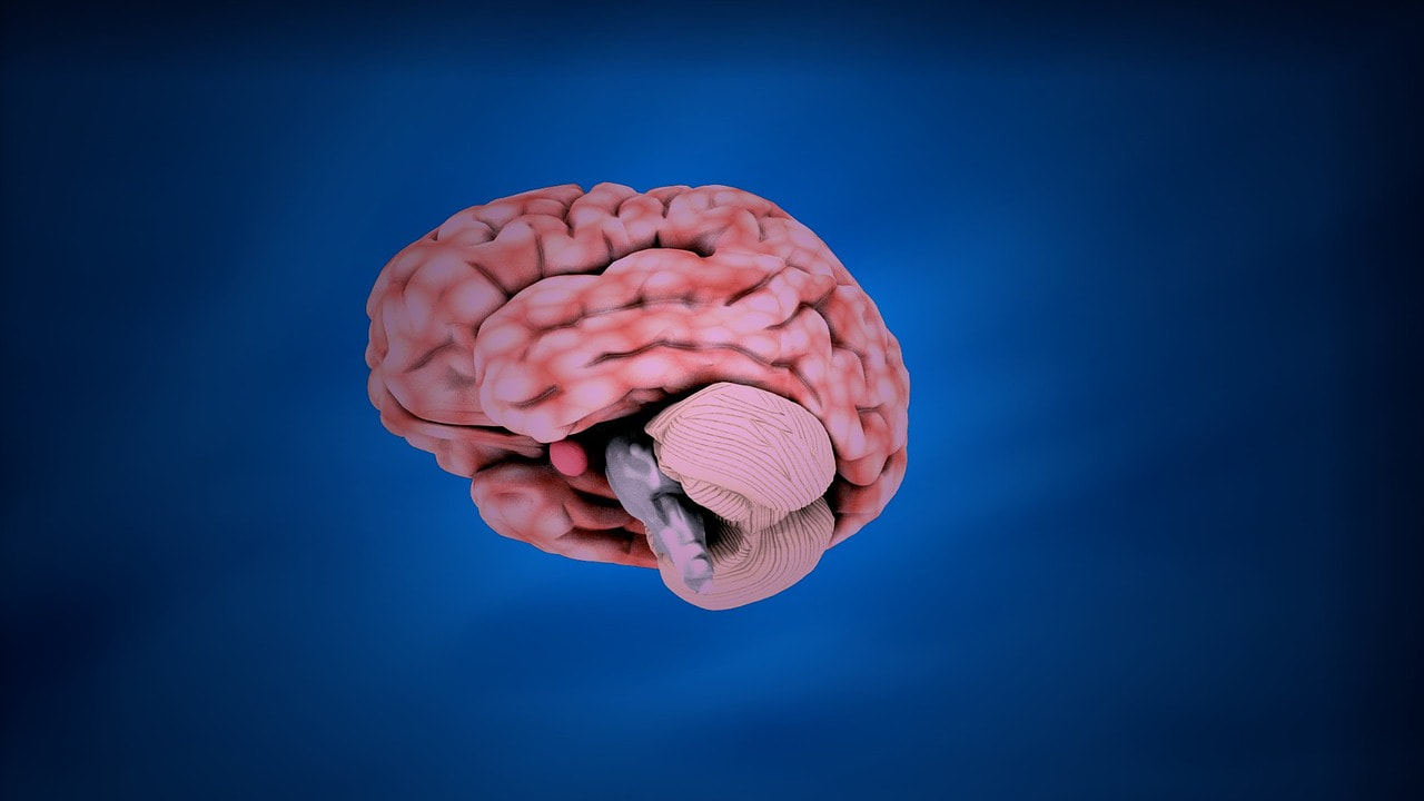
Cerebellum is a Latin word that means ‘little brain’. This term refers to its distinctive appearance compared to the rest of the brain. The cerebellum is a wrinkled, cauliflower-shaped brain structure located at the base of the skull. It looks like it could be a second brain and in fact, it is in some ways. For example, it has more total neurons than every other part of the brain combined. These neurons are organized in a way that creates tremendous processing power which is also a specific feature of the cerebellum. Though on the surface it seems like the cerebellum could operate just fine on its own, it is very much one part of a whole. It’s highly connected with many other areas of the brain and serves an important role in a variety of brain functions, most notably motor control.
Location of the Cerebellum
The cerebellum is located at the base of the skull, in the area where the brain and spinal cord meet. With respect to development, the cerebellum is in an area known as the hindbrain. This region contains the evolutionarily oldest parts of the brain including regions that control the most basic life functions such as breathing.

Anatomy of the Cerebellum
The structure of the cerebellum is really quite exquisite. It’s complex yet remarkably orderly, and if you ever have the pleasure of looking at cerebellar tissue under a microscope, you’ll notice that the cell types that comprise it are strikingly beautiful. Fully describing the anatomical organization of the cerebellum could fill up an entire book, so we’ll just stick with the basics.
First, let’s start with the anatomical structures we can see with the naked eye. If you were to look at a cerebellum, you would notice that it is divided into two hemispheres connected by a central structure called the vermis (Latin for worm). The surface of the cerebellar hemispheres is characterized by a series of ridges or folds, called folia, which increase the surface area of the cerebellum and allow for greater numbers of neurons. These folia are separated by deep grooves, called fissures, which divide the cerebellum into several lobes (Kandel et al., 2000).
The cerebellum can be divided into three main lobes: the anterior lobe, the posterior lobe, and the flocculonodular lobe. The anterior lobe is the largest and is responsible for controlling movements of the limbs and body. The posterior lobe is involved in the coordination of movements and receives input from the cerebral cortex. The flocculonodular lobe is located at the bottom of the cerebellum and is involved in the coordination of eye movements (Kandel et al., 2000).
Cerebellar anatomy at the cellular level is widely regarded as exceptionally beautiful. With respect to this level of observation, the cerebellum is made up of three main layers: the molecular layer, the Purkinje cell layer, and the granular layer (Kandel et al., 2000).
The Molecular Layer
The molecular layer is the outermost layer of the cerebellum and is made up of small neurons, called interneurons, as well as the axons and dendrites of the Purkinje cells.
The Purkinje Cell Layer
The Purkinje cell layer is located in the middle of the cerebellum and consists of large, flat neurons called Purkinje cells. The axons of the Purkinje cells extend into the molecular layer, where they form synapses with other neurons.
The Granular Layer
The granular layer is the innermost layer of the cerebellum and is composed of small, densely packed neurons called granule cells. The axons of the granule cells extend into the molecular layer, where they form parallel fibers that run perpendicular to the Purkinje cells. The parallel fibers form synapses with the dendrites of the Purkinje cells and other interneurons in the molecular layer.
Functions of the Cerebellum
- Temporal prediction
- Balance
- Proprioception
- Motor coordination
- Spatial orientation
- Generating large force at movement execution
- Motor planning
- Posture
- Speech
Cerebellum vs Cerebrum
The cerebrum is 90% of the total brain volume and is responsible for a wide range of functions, including sensory processing, perception, thought, and memory. This part of the brain is also responsible for higher cognitive functions such as consciousness, decision-making, problem-solving, and language.
Cerebellum vs Basal Ganglia
As mentioned previously, the cerebellum sits at the base of the brain and is involved in coordinating movement, maintaining balance, and fine-tuning motor actions. It receives input from the sensory systems, motor cortex, and brainstem, and sends output to the motor cortex and brainstem to control muscle tone, movement speed, and accuracy.
The basal ganglia, on the other hand, are a group of nuclei located deep in the brain, consisting of the striatum, globus pallidus, substantia nigra, and subthalamic nucleus. They are involved in motor planning and execution, as well as reward processing, habit formation, and emotion regulation. They receive input from the cortex and thalamus and send output to the motor cortex and brainstem to initiate and modulate movement.

The Role of the Cerebellum in Balance
The cerebellum plays a crucial role in maintaining balance and coordination of movements. It receives input from sensory organs such as the inner ear, visual system, and proprioceptive system (which detects the position and movement of body parts), and integrates this information to coordinate and adjust body movements (Morton & Bastian, 2004).
The cerebellum uses this sensory input to generate motor commands that adjust the activity of muscles, tendons, and joints to maintain posture and balance. For example, when a person tilts forward, the cerebellum sends signals to the muscles in the legs and back to adjust their activity and prevent the person from falling.
The cerebellum also works in conjunction with other parts of the brain, such as the vestibular system and the brainstem, to control balance and posture. Together, these systems enable us to maintain balance and stability while standing, walking, or performing other movements (Barmack & Pettorossi, 2021).
The Role of the Cerebellum in Memory
Working Memory
Working memory is generally defined as the ability to hold and manipulate a small amount of information. This capacity facilitates comprehension, problem-solving, reasoning, and planning.
Implicit Memory
Implicit memories are unconscious and often unintentionally created. Learning a new skill such as roller skating is an example of implicit memory.
Explicit Memory
Explicit memory is basically the opposite of implicit memory. These are memories that are created consciously and that can be described verbally. A memory of an important event is an example of explicit memory.
Damage to the Cerebellum: Ataxia
Cerebellar ataxia is a neurological disorder that is characterized by a lack of muscle coordination, which can result in unsteady gait, difficulty with fine motor tasks, as well as speech and eye movement abnormalities. This disorder can be caused by several different factors, including genetic mutations, autoimmune disorders, infections, toxins, trauma, and tumors (Marsden & Harris, 2011).
Symptoms of cerebellar ataxia can vary depending on the underlying cause and the severity of the condition. Common symptoms include unsteady gait, tremors, difficulty with fine motor tasks such as writing, and slurred or slow speech. In more severe cases, individuals with cerebellar ataxia may also experience difficulty swallowing, vision problems, and cognitive impairment (Marsden, 2018).
There is no cure for cerebellar ataxia but treatment options are available to help manage symptoms and improve quality of life. Treatments may include physical therapy to improve balance and coordination, medications to manage tremors or other symptoms, and assistive devices to help with mobility and daily tasks.
Hypoplasia in the Cerebellum
Hypoplasia is a medical term that refers to the underdevelopment or incomplete development of a tissue or organ. In the context of the cerebellum, hypoplasia refers to a condition where the cerebellum is smaller than normal or has fewer cells than it should (Poretti et al., 2014).
Cerebellar hypoplasia can occur as a result of various genetic, environmental, or developmental factors. It can be present at birth (congenital) or develop later in life. Some causes of cerebellar hypoplasia include genetic disorders, such as Dandy-Walker syndrome and Joubert syndrome, exposure to certain toxins during pregnancy, infections such as rubella or cytomegalovirus, and traumatic brain injury (Poretti et al., 2014).
Stroke in the Cerebellum
- Ataxia
- Nausea and vomiting
- Headache
- Dizziness and vertigo
- Difficulty speaking
- Difficulty with eye movements and double vision
- Deficits in attention and working
The severity and duration of these symptoms may vary depending on the extent of the damage and the location of the stroke in the cerebellum. Rehabilitation, physical therapy, and medication may be used to manage symptoms and improve the quality of life for individuals who suffered a cerebellar stroke.
The Cerebellum and White Matter
White matter in the cerebellum consists of myelinated axons, which are long, thin structures that transmit electrical impulses between neurons. Myelin is a fatty substance that wraps around these axons, allowing for faster transmission of impulses.
The white matter in the cerebellum is organized into a complex network of pathways that allow for the coordination of movement, balance, and other motor functions. The white matter in the cerebellum plays a critical role in connecting different regions of the cerebellum and transmitting information between the cerebellum and other parts of the brain.

Articles Related to The Cerebellum
Books Related to The Cerebellum
Final Thoughts on the Cerebellum
The cerebellum is a unique and interesting brain structure with a plethora of important functions. It supports fine motor control, motor timing, walking, speech, attention, posture, spatial orientation, balance, and motor planning – just to name a few. It also has a highly organized structure and immense processing power. It comprises more neurons than all other brain regions combined even though it only occupies 10% of the total brain volume. If you would like to learn more about the wonders of the cerebellum, check out this video:
Video: The Cerebellum
Don’t Forget to Grab Our Free eBook to Learn How to
Grow Your Wellness Business Exponentially!
References
- Barmack, N. H., & Pettorossi, V. E. (2021). Adaptive balance in posterior cerebellum. Frontiers in Neurology, 12, 635259.
- Kandel, E. R., Schwartz, J. H., Jessell, T. M., Siegelbaum, S., Hudspeth, A. J., & Mack, S. (Eds.). (2000). Principles of neural science (Vol. 4, pp. 833-850). New York: McGraw-hill.
- Marsden, J. F. (2018). Cerebellar ataxia. Handbook of clinical neurology, 159, 261-281.
- Marsden, J., & Harris, C. (2011). Cerebellar ataxia: pathophysiology and rehabilitation. Clinical rehabilitation, 25(3), 195-216.
- Morton, S. M., & Bastian, A. J. (2004). Cerebellar control of balance and locomotion. The neuroscientist, 10(3), 247-259.
- Poretti, A., Boltshauser, E., & Doherty, D. (2014, June). Cerebellar hypoplasia: differential diagnosis and diagnostic approach. In American Journal of Medical Genetics Part C: Seminars in Medical Genetics (Vol. 166, No. 2, pp. 211-226).
- Rapoport, M., van Reekum, R., & Mayberg, H. (2000). The role of the cerebellum in cognition and behavior: a selective review. The Journal of neuropsychiatry and clinical neurosciences, 12(2), 193-198.
- Stoodley, C. J., MacMore, J. P., Makris, N., Sherman, J. C., & Schmahmann, J. D. (2016). Location of lesion determines motor vs. cognitive consequences in patients with cerebellar stroke. NeuroImage: Clinical, 12, 765-775.
Are You a Therapist, Coach, or Wellness Entrepreneur?
Grab Our Free eBook to Learn How to Grow Your Wellness Business Fast!